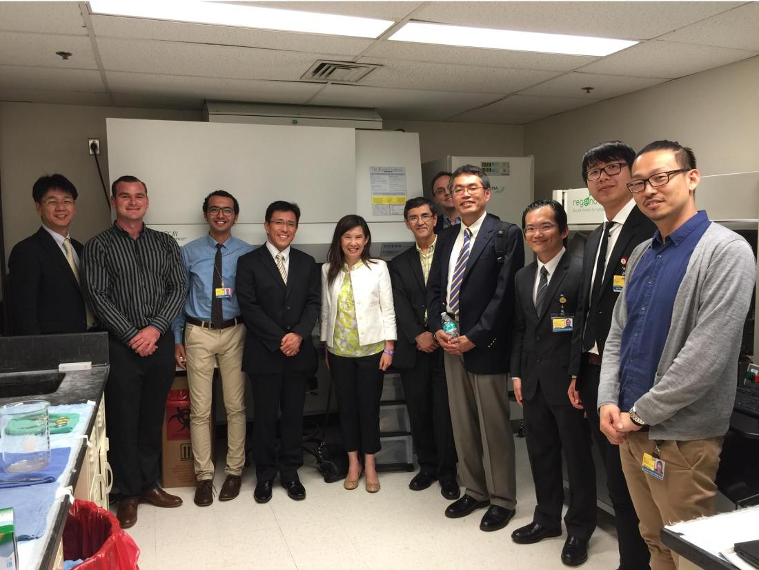
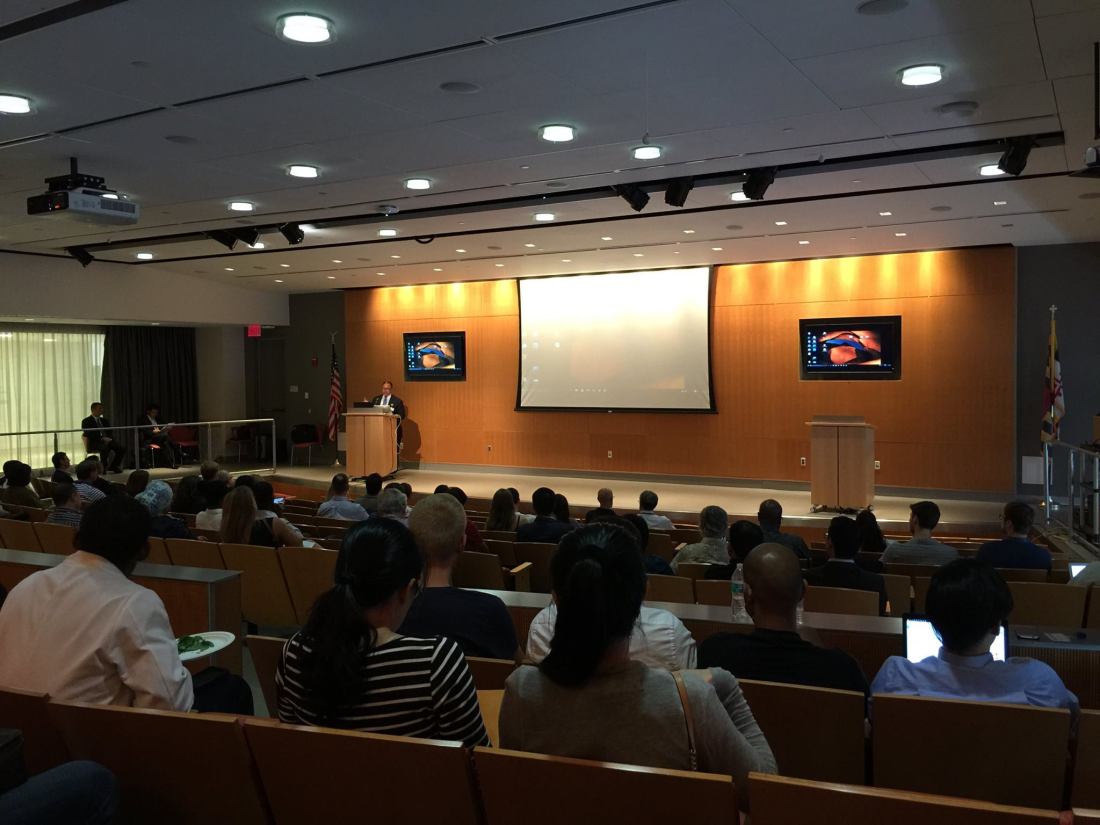
1st Scaffold-free 3D Bioprinting Seminar (2 Sept 2016)
Thank you everyone for coming! In case you missed the seminar, here is the webcast link.


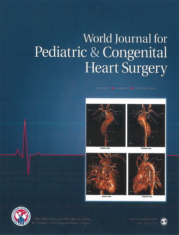
Loeys-Dietz syndrome (LDS) is an autosomal dominant genetic connective tissue disorder associated with aortic aneurysmal disease. Kommerell diverticulum (KD) is a rare aortic diverticulum, for which the indication for surgery and the surgical techniques remain subjects of debate. We describe our experience with a successful total aortic arch replacement including KD resection through a median sternotomy for a pediatric patient with LDS.
© The Author(s) 2016.
Kommerell diverticulum; Loeys-Dietz syndrome; aberrant right subclavian artery; congenital heart disease
Lab Meeting 09/03/2016
Discussed the successful 3D Bioprinting symposium yesterday, attendance 200+.
New members, welcome!
Sam Hardy
Ananya Gupta
Gunnar Mattson
Projects discussed:
Cardiac 3D Tissue Printing Project
TG II in vitro study
EVC (Early Vascular Cells)
Spheroid review
TEVG studies
Presented papers:
1. Şenel Ayaz HG, Perets A, Ayaz H, Gilroy KD, Govindaraj M, Brookstein D, Lelkes PI. Textile-templated electrospun anisotropic scaffolds for regenerative cardiac tissue engineering. Biomaterials. 2014 Oct;35(30):8540-52. doi: 10.1016/j.biomaterials.2014.06.029. Epub 2014 Jul 10.
2. Henry E1, Cores J1, Hensley MT2, Anthony S3, Vandergriff A1, de Andrade JB2, Allen T2, Caranasos TG4, Lobo LJ5, Cheng K6. Adult Lung Spheroid Cells Contain Progenitor Cells and Mediate Regeneration in Rodents With Bleomycin-Induced Pulmonary Fibrosis. Stem Cells Transl Med. 2015 Nov;4(11):1265-74. doi: 10.5966/sctm.2015-0062. Epub 2015 Sep 10.
3. Krawiec JT1,2, Weinbaum JS1,2, Liao HT3,4, Ramaswamy AK1,2, Pezzone DJ1, Josowitz AD1, D’Amore A1,2,5,6, Rubin JP2,3, Wagner WR1,2,5,7, Vorp DA1,2,5,7,8. In Vivo Functional Evaluation of Tissue-Engineered Vascular Grafts Fabricated Using Human Adipose-Derived Stem Cells from High Cardiovascular Risk Populations. Tissue Eng Part A. 2016 May;22(9-10):765-75. doi: 10.1089/ten.TEA.2015.0379.
4. Amaral AJ1, Pasparakis G1. Rapid Formation of Cell Aggregates and Spheroids Induced by a “Smart” Boronic Acid Copolymer. ACS Appl Mater Interfaces. 2016 Aug 29. [Epub ahead of print]
5. Dawson KA1, Yan Y1. Drug delivery: Leukocyte-like carriers. Nat Mater. 2016 Aug 24;15(9):935-6. doi: 10.1038/nmat4737.
IncuCyte ZOOM™ Continuous Live-cell Imaging & Analysis System
http://www.selectscience.net/products/incucyte-zoom-continuous-live-cell-imaging-and-analysis-system/?prodID=116928
Corning® stemgro® hMSC Medium
http://cellgro.com/products/new-products/corning-stemgro-hmsc-medium.html
Note (5/3/2017): Some time after this post, lab meeting summaries and news updates were shifted to Facebook, under http://www.facebook.com/hibinolab/.
Launch of the Hibinolab Facebook Page: https://www.facebook.com/hibinolab
Please feel free to check it out!
Projects discussed:
Articles discussed / Journal Club:
Tissue engineered vascular grafts (TEVGs) have the potential to overcome the issues faced by existing small diameter prosthetic grafts by providing a biodegradable scaffold where the patient’s own cells can engraft and form functional neotissue. However, applying classical approaches to create arterial TEVGs using slow degrading materials with supraphysiological mechanical properties, typically results in limited host cell infiltration, poor remodeling, stenosis, and calcification. The purpose of this study is to evaluate the feasibility of novel small diameter arterial TEVGs created using fast degrading material. A 1.0mm and 5.0mm diameter TEVGs were fabricated with electrospun polycaprolactone (PCL) and chitosan (CS) blend nanofibers. The 1.0mm TEVGs were implanted in mice (n = 3) as an unseeded infrarenal abdominal aorta interposition conduit., The 5.0mm TEVGs were implanted in sheep (n = 6) as an unseeded carotid artery (CA) interposition conduit. Mice were followed with ultrasound and sacrificed at 6 months. All 1.0mm TEVGs remained patent without evidence of thrombosis or aneurysm formation. Based on small animal outcomes, sheep were followed with ultrasound and sacrificed at 6 months for histological and mechanical analysis. There was no aneurysm formation or calcification in the TEVGs. 4 out of 6 grafts (67%) were patent. After 6 months in vivo, 9.1 ± 5.4% remained of the original scaffold. Histological analysis of patent grafts demonstrated deposition of extracellular matrix constituents including elastin and collagen production, as well as endothelialization and organized contractile smooth muscle cells, similar to that of native CA. The mechanical properties of TEVGs were comparable to native CA. There was a significant positive correlation between TEVG wall thickness and CD68+ macrophage infiltration into the scaffold (R2 = 0.95, p = 0.001). The fast degradation of CS in our novel TEVG promoted excellent cellular infiltration and neotissue formation without calcification or aneurysm. Modulating host macrophage infiltration into the scaffold is a key to reducing excessive neotissue formation and stenosis.

